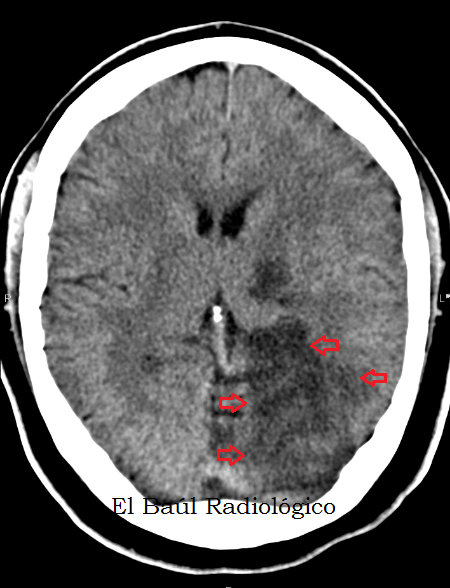
Cuando se produce la oclusión brusca del flujo sanguíneo en una arteria del encéfalo, del hígado o del riñón, por ejemplo, el parénquima irrigado por esa arteria se muere y el resultado final es un infarto. En el hígado o en el riñón, la zona infartada cicatriza al cabo del tiempo y se recupera la función de ambas vísceras con toda normalidad. En el cerebro no sucede así, porque las funciones de relación se agrupan en "áreas motoras y sensitivas" que tienen una distribución topográfica muy definida en el cerebro. Por eso cuando se produce un infarto cerebral y se mueren las neuronas responsables de una función determinada, como el habla o la visión, el déficit ya no se recuperará con el paso del tiempo como sucede con los infartos hepáticos o renales. En las siguientes imágenes presentamos un caso de POCI subagudo izquierdo (24 horas). La obstrucción ha afectado al tronco de la arteria cerebral posterior izquierda y esa circunstancia, nos permite apreciar con total precisión, el territorio irrigado por dicha arteria y predecir las secuelas que padecerá esa persona, un varón de 65 años.
(In the pictures below, wepresent a case of a subacuteleftPOCI(24hours). The obstructionhas affected thetrunk of theleftposterior cerebralarteryand this circumstanceallows us to appreciate, with precision, the territorysupplied bythis arteryand predictthe sequels thatthepatientis going to suffer)
(In the pictures below, wepresent a case of a subacuteleftPOCI(24hours). The obstructionhas affected thetrunk of theleftposterior cerebralarteryand this circumstanceallows us to appreciate, with precision, the territorysupplied bythis arteryand predictthe sequels thatthepatientis going to suffer)
FIGURA 1) Angiografía por Resonancia Magnética, con Reconstrucción de Máxima Intensidad (MIP) de las principales arterias que constituyen el Polígono de Willis. Representación aproximada del territorio irrigado por la arteria Cerebral Posterior Izquierda.
(MagneticResonance Angiography (MRA) withMaximum IntensityProyection(MIP) of the mainarteries thatform theCircle of Willis. Approximate representationof theterritory irrigated by theLeftPosterior CerebralArtery)
FIGURA 2) Pequeña área hipodensa (flechas) que representa un infarto isquémico subagudo en el lóbulo temporal izquierdo.
(Smallhypodense area (arrows)that represents theischemic infarctionin theleft temporallobe)
FIGURA 3) Siguiente corte más cefálico. Area hipodensa (flechas), circunscrita, que representa el infarto isquémico. Se aprecia con claridad cómo respeta el hemisferio izquierdo del cerebelo, irrigado por la arteria cerebelosa postero-superior izquierda.
(A morecephalad nextslice.Hypodensearea(arrows), circumscribed, representingischemic stroke.It isclearly seenhow isrespectedtheleft cerebellar hemisphere,supplied by the leftposterior superiorcerebellar artery)
FIGURA 4) En la zona infartada se ha producido la muerte de las células: gliales, neuronas y las que forman el endotelio capilar. Eso provoca la extravasación de agua desde los capilares lesionados y del citoplasma de las células. El resultado es un tejido muerto "encharcado" de agua. Por eso adquiere esa tonalidad oscura y presenta unos valores de atenuación más bajos que el tejido normal: Infarto isquémico subagudo (hipodenso 16 UH).
(The infarcted areahasresulted in deathofcells: glial cells,neurons and capillary endothelium. This, causeswaterextravasationfrom damagedcapillaries and from the cytoplasmof cells.The result is adead tissue"puddled" of water.Sothat, the infarcted áreabecomesdarkand hasalowerattenuation valuesthan normal tissue:Subacuteischemicinfarction)
FIGURA 5) Infarto isquémico subagudo: POCI izquierdo.
(Subacuteischemicinfarction:leftPOCI)
FIGURA 6) Infarto isquémico subagudo: POCI izquierdo. El área hipodensa delimita perfectamente el territorio de la arteria cerebral posterior izquierda.
(Subacuteischemicinfarction:leftPOCI.Thehypodense areadelineatesperfectlythe territory of theleftposterior cerebralartery.
FIGURA 7) Infarto isquémico subagudo: POCI izquierdo. El área hipodensa delimita el territorio de la arteria cerebral posterior izquierda.
(Subacuteischemicinfarction: leftPOCI. Thehypodense areadelineates the territory of theleftposterior cerebralartery.
FIGURA 8) Infarto isquémico subagudo: POCI izquierdo. A medida que se realizan cortes más cefálicos, el área hipodensa disminuye de tamaño.
(Subacuteischemicinfarction:leftPOCI)Asslicesare performed more cephalad, the hypodenseareadecreases in size)
FIGURA 9) En el último corte apenas es perceptible el infarto, pero en toda la secuencia de imágenes que se han mostrado hemos podido apreciar el territorio exacto irrigado por la arteria cerebral posterior izquierda y el resultado de un POCI secundario a la oclusión de la rama principal de la arteria.
(Inthe last sliceis hardly noticeable the infart area,butthe entire sequenceof images, thatwe have seen,has showntheexact territorysupplied by theleftposterior cerebralarteryandthe result of aPOCIaffectingthe main branch ofthis artery.)
Hospital Universitario Miguel Servet (HUMS) Zaragoza. Spaiñ


![An American Tail [1986] [DVD5-R1] [Latino]](http://iili.io/FjktrS2.jpg)


