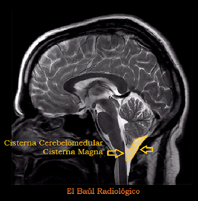
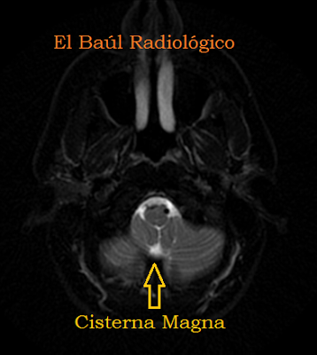
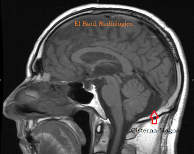
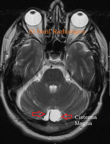
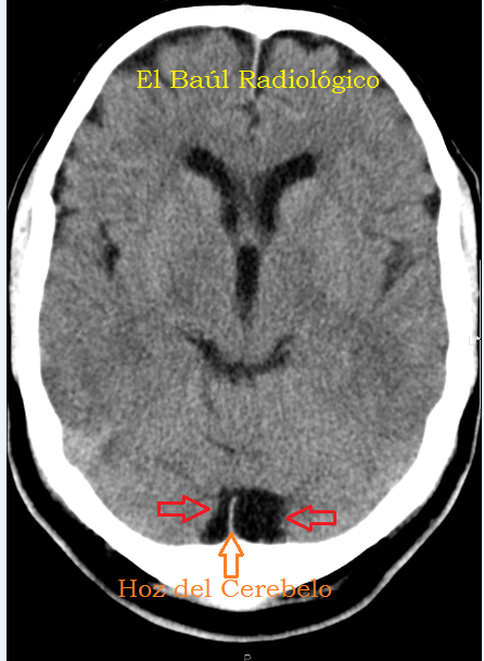
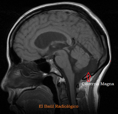
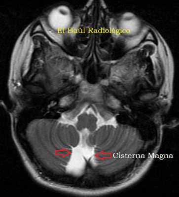
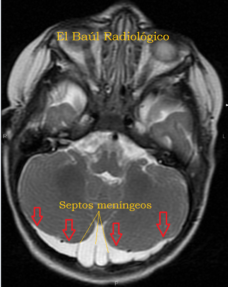
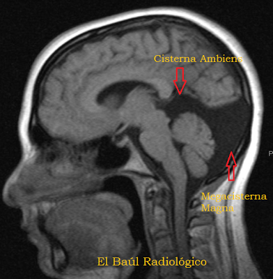
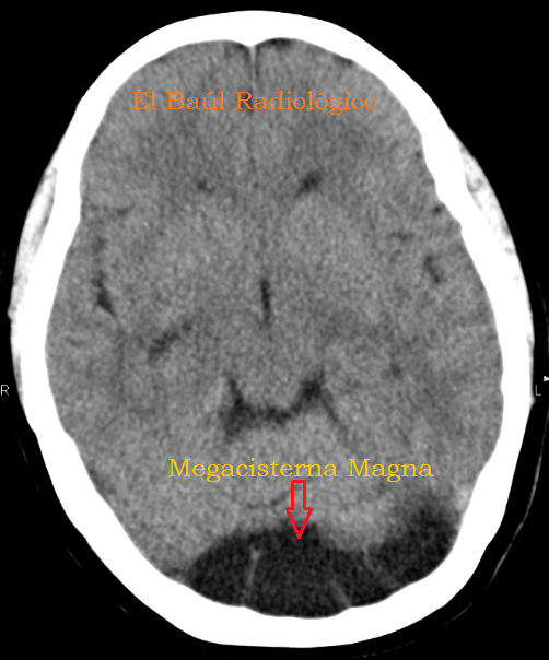
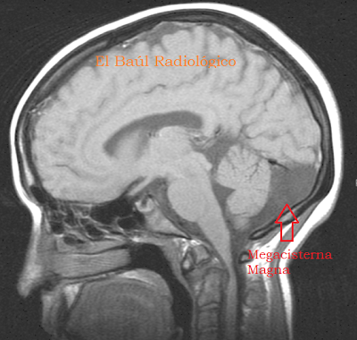
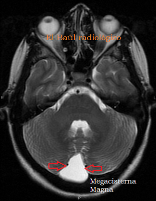
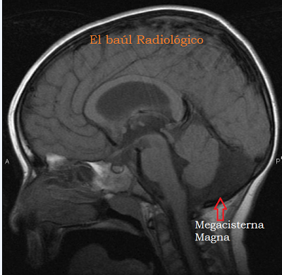
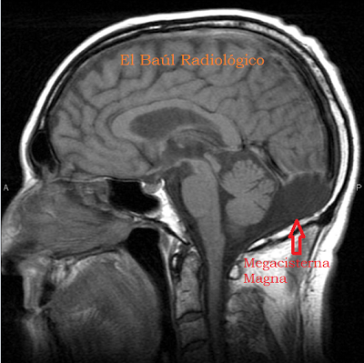
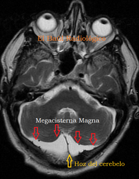
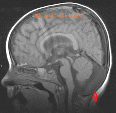
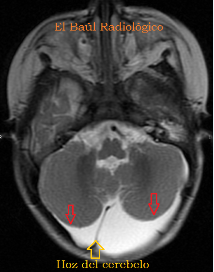
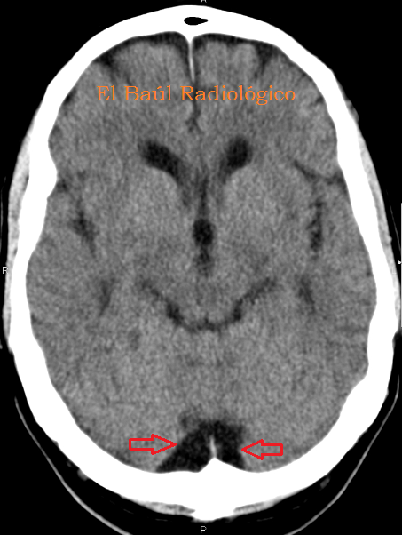
Con el nombre de Cisterna Magna o Cerebelomedular (figuras 1, 2, 3 y 4), se conoce a una parte del espacio subaracnoideo de la fosa posterior craneal donde drena el líquido cefalorraquídeo desde el IV ventrículo, a traves de los agujeros de Luschka laterales y el central de Magendie. No tiene mucha importancia desde el punto de vista anatómico, pero su morfología y el tamaño son tan variables que inducen a confusión, cuando se aprecia de mayor tamaño que el esperado. Cuando este espacio es mas grande de lo normal se denomina Megacisterna Magna. Es una variante anatómica que no tiene significación patológica. El aumento de volumen de la cisterna es secundario a un defecto congénito en el desarrollo del cerebelo, especialmente el vermis. Nunca debe confundirse con un quiste aracnoideo que sí podría tener consecuencias patológicas. A continuación presentamos varios casos de cisternas magnas, descubiertas de manera fortuita en exploraciones de TC y TRM realizadas por problemas neurológicos que no tenían relación con el tamaño de la cisterna.
(Withthe nameofCisternaMagnaor Cerebellomedullary Cistern (Figures 1,2,3 and 4), is knownto a part ofsubarachnoid space of theposteriorcranialfossa, wherecerebrospinal fluid drainsfrom thefourth ventriclethroughthelateralholes ofLuschkaand the central of Magendie. Itis not very importantfrom the point ofanatomical view, but their morphologyand sizevariablesare soconfusingwhenwe appreciate itlargerthan expected.Whenthis spaceis biggerthan normalis calledMega Cisterna Magna. It is anormalanatomic variantwithoutpathological significance. The increasedvolumeofthe cistern isdueto a congenital defectinthedevelopmentof the cerebellum,especiallythevermis. It wil neverto be confused withan arachnoid cyst. Here areseveral cases ofcisternsmagna, discovered fortuitouslyinCT andMRTexamsperformed byneurological problems thatwere not related tothe sizeof the cisterns).
FIGURA 1) TRM sagital. La Cisterna Magna está delimitada por la superficie inferior del cerebelo, la cara posterior de la médula cervical y la superficie interna del hueso occipital, recubierto éste por la duramadre y la aracnoides.
(MRI saggital.The CisternMagnais delimitedbythelowersurfaceof the cerebellum,theposteriorfaceofthe cervical cordandtheinner surface of theoccipitalbonecoveredbythearachnoidanddura mater membranes).
FIGURA 2) Vista de la Cisterna Magna en proyección coronal.(View of theCisternaMagnaincoronalprojection)
FIGURA 3) Vista de la Cisterna Magna en proyección axial.
(View of theCisternaMagnainaxialprojection)
FIGURA 4) En la mayoría de las personas la Cisterna Magna es tan pequeña que apenas es visible.
(Inmany people theCisternaMagnais so smallthat it is hardlyvisible).
CASO 1
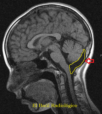
FIGURA 1) El tamaño y la morfología de la Cisterna Magna son congénitos, por eso ya puede verse más grande de lo habitual, en exploraciones de TRM realizadas en niños. Flecha roja.
(The sizeand morphologyof theCisternaMagnaare congenital. So that's the causeofit is foundlargerthan usual in MRIscansperformed in children. Red arrow)
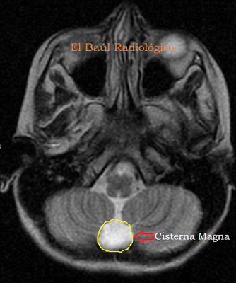
FIGURA 2) Vista de la Cisterna Magnanormal en proyección axial.
(View of a normalCisternaMagnainaxialprojection)
CASO 2
FIGURA 1)Vista de la Cisterna Magnanormal en proyección sagital.
(View of a normalCisternaMagnainsaggitalprojection)
FIGURA 2)Vista de la Cisterna Magnanormal en proyección axial. La existencia de la hoz del cerebelo permite distinguirla de un quiste aracnoideo.
(View of a normalCisternaMagnainaxialprojection.The existence ofthe falx cerebellicandistinguish it froman arachnoid cyst.)
FIGURA 3) Vista de la Cisterna Magnanormal en una imagen de TC axial. La existencia de la hoz del cerebelo permite distinguirla de un quiste aracnoideo.
(View of a normalCisternaMagnain a TCaxialprojection.The existence ofthe falx cerebellicandistinguish it froman arachnoid cyst.)
CASO 3
FIGURA 1) Vista de la Cisterna Magna más grande de lo habitual. ¿Megacisterna Magna?. Normal.
(Sagittal viewof theCisternaMagnalargerthan usual. Megacisterna Magna?. Normal )
FIGURA 2) Megacisterna Magna, normal.
Megacisterna Magna, normal )
CASO 4
FIGURA 1) Vista de la Cisterna Magna más grande de lo habitual. Megacisterna Magna, normal.
(Sagittal viewof theCisternaMagnalargerthan usual. Megacisterna Magna, normal )
FIGURA 2) Megacisterna Magna en una imagen de TRM axial. La existencia de septos meníngeos, y las extensiones laterales en forma de alas, permite distinguirla de un quiste aracnoideo.
(View of the MegacisternaMagna in this MRI axial image. The existenceofmeningealsepta, andthe lateral extensionsin formof wings, enables us to distinguish it from anarachnoidcyst)
(View of the MegacisternaMagna in this MRI axial image. The existenceofmeningealsepta, andthe lateral extensionsin formof wings, enables us to distinguish it from anarachnoidcyst)
CASO 5
FIGURA 1) Vista en proyección sagital de una Megacisterna Magna.También aparece aumentada de tamaño la cisterna ambiens. Atrofia vermiana.
(Sagittalprojectionof aMegacisternaMagna. Alsoappearsenlargedthe Ambient Cistern. Vermianatrophy.)
FIGURA 2) Megacisterna Magna en una imagen de TRM axial. La existencia de septos meníngeos, y las extensiones laterales en forma de alas, permite distinguirla de un quiste aracnoideo.
(MegacisternaMagna in this MRI axial image. The existenceofmeningealsepta, andthe lateral extensionsin formof wings, enables us to distinguish it from anarachnoidcyst)
(MegacisternaMagna in this MRI axial image. The existenceofmeningealsepta, andthe lateral extensionsin formof wings, enables us to distinguish it from anarachnoidcyst)
FIGURA 3) Megacisterna Magna en una imagen de TC axial.
(MegacisternaMagna in this CT axial image.)
CASO 6
FIGURA 1) Proyección sagital de una Megacisterna Magna. Atrofia vermiana.
(Sagittalprojectionof aMegacisternaMagna.Vermianatrophy.)
FIGURA 2) Megacisterna Magna en una imagen de TRM axial. Atrofia Vermiana
(MegacisternaMagna in this MRI axial image. Vermian Atrophy)
(MegacisternaMagna in this MRI axial image. Vermian Atrophy)
CASO 7
FIGURA 1) Proyección sagital de una Megacisterna Magna. Atrofia vermiana.
(Sagittalprojectionof aMegacisternaMagna.Vermianatrophy.)
FIGURA 2) Megacisterna Magna en una imagen de TRM axial. Se aprecia la Hoz del cerebelo que la parte en dos. Atrofia cerebelosa.
(MegacisternaMagna in this MRI axial image. Cerebellar atrophy. The falx cerebelli)
(MegacisternaMagna in this MRI axial image. Cerebellar atrophy. The falx cerebelli)
CASO 8
FIGURA 1) Proyección sagital de una Megacisterna Magna. Atrofia vermiana.
(Sagittalprojectionof aMegacisternaMagna.Vermianatrophy.)
FIGURA 2) Megacisterna Magna en una imagen de TRM axial.Atrofia cerebelosa. Hoz del cerebelo.
(MegacisternaMagna in this MRI axial image. Cerebellar atrophy. Falx cerebelli)
(MegacisternaMagna in this MRI axial image. Cerebellar atrophy. Falx cerebelli)
CASO 9
FIGURA 1) Proyección sagital de una Megacisterna Magna. Atrofia vermiana.
(Sagittalprojectionof aMegacisternaMagna.Vermianatrophy.)
FIGURA 2) Megacisterna Magna en una imagen de TRM axial.Hoz del cerebelo. Atrofia cerebelosa.
(MegacisternaMagna in this MRI axial image. Falx cerebelli. Cerebellar atrophy)
(MegacisternaMagna in this MRI axial image. Falx cerebelli. Cerebellar atrophy)
CASO 10
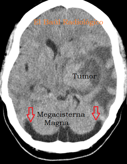
FIGURA 1) Megacisterna Magna en una imagen de TC axial.
(MegacisternaMagna in this CT axial image. )
(MegacisternaMagna in this CT axial image. )
FIGURA 2) Megacisterna Magna en una imagen de TC axial. Hoz del cerebelo.
(MegacisternaMagna in this CT axial image. Falx cerebelli )
(MegacisternaMagna in this CT axial image. Falx cerebelli )
CASO 11
FIGURA 1) Megacisterna Magna en una imagen de TC axial. Septos meningeos. Atrofia cerebelosa
(MegacisternaMagna in this CT axial image. Meningealsepta. Cerebellar atrophy.)
(MegacisternaMagna in this CT axial image. Meningealsepta. Cerebellar atrophy.)
FIGURA 2) Megacisterna Magna. Septos meningeos. Atrofia cerebelosa
(MegacisternaMagna. Meningealsepta. Cerebellar atrophy.)
Hospital Universitario Miguel Servet (HUMS) Zaragoza. Spaiñ
